A multimodal analysis of Magnetic Resonance Imaging for the study of brain abnormalities in migraine: gray matter morphometry, white matter integrity and structural connectivity

A multimodal analysis of Magnetic Resonance Imaging for the study of brain abnormalities in migraine: gray matter morphometry, white matter integrity and structural connectivity

PhD in Information Technology and Telecommunications
Doctor: Álvaro Planchuelo Gómez
Date of reading: 01/06/2021
Directors: Santiago Aja Fernández, Rodrigo de Luis García
Court: Silvia de Santis (chairwoman), Juan Pablo Casaseca de la Higuera (secretary), Julio Pascual Gómez (member)

Introduction

Migraine is a primary disease characterized by recurrent headache attacks. Despite the high migraine prevalence and its consequences, currently there are no migraine biomarkers. Furthermore, the migraine pathophysiology is not completely understood yet. In order to find a migraine biomarker and better understand the migraine pathophysiology, Magnetic Resonance Imaging (MRI) has been employed.

Objectives

The main objective was the characterization of gray matter and white matter structural properties of patients with Chronic Migraine (CM) and Episodic Migraine (EM), the two types currently distinguished. The use of advanced and novel diffusion measures not employed previously in the migraine literature was considered to provide an additional strategy for the assessment of white matter changes. Moreover, the structural connections between gray matter regions through the white matter tracts were assessed, bearing in mind their possible relationship with gray matter morphometry.

Development

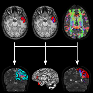
MATERIALS AND METHODS: Morphometry measures for gray matter and diffusion tensor imaging (DTI) parameters in combination with tract-based spatial statistics (TBSS) for white matter were used to assess migraine patients together with more advanced techniques such as tractography and connectomics. Moreover, novel methods recently developed such as Apparent Measures Using Reduced Acquisitions (AMURA) to evaluate diffusion properties were applied. To analyze the relationship between the changes in diverse tissues, the fusion method multimodal Canonical Correlation Analysis followed by joint Independent Component Analysis (mCCA-jICA) was used.

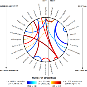
RESULTS: The sample of the assessments with diffusion MRI data included 56 CM patients, 54 EM patients and 50 healthy controls (HC), with an additional subject per group for the analysis of gray matter morphometry. In the morphometry comparisons, higher cortical curvature values and lower cortical thickness, gray matter volume and surface area values were found in migraine patients (one or both groups) compared to HC. Furthermore, surface area differences between both migraine groups were identified, with lower values in CM. Regarding the white matter analysis with the DTI descriptors, different trends were obtained in CM with respect to HC. Higher and lower fractional anisotropy, and lower radial diffusivity were found in CM compared to HC. Higher axial diffusivity in EM compared to HC was identified. With AMURA, additional differences between EM and HC that could not be identified with conventional DTI parameters were found. Lower return-to-origin probability values were observed in EM compared to HC. In addition, lower axial diffusivity values in CM compared to EM were found, obtaining similar results with AMURA and other DTI parameters. Regarding connectomics, debilitated structural connections between regions within the diverse lobes and strengthened connections with pain processing regions were found. Weakened structural connectivity in CM compared to EM was detected connections between the caudate nucleus and the orbitofrontal cortex, while strengthened specific altered connectivity was found in connections with the hippocampus in CM patients. The changes of structural connectivity were associated with cortical curvature alterations.

Conclusions

Migraine (CM and EM) is characterized by a series of structural gray and white matter changes, including connections between specific gray matter regions. Considering the comparison between both migraine groups, CM follows a set of alterations different from the changes between EM and HC. The results suggest that CM is an entity with unique properties and substantially different from EM. The surface area could work as a CM biomarker. AMURA can be employed to complement the results of the classical DTI approach, even in suboptimal conditions to exploit the technique. The use of sophisticated fusion approaches such as mCCA-jICA allows the identification of the relationship between the changes observed in diverse MRI modalities.